Sonography/Ultrasound (US)
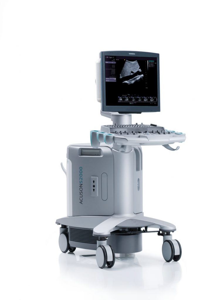
Sonography, also called echography or ultrasound, is the most widely used imaging method in healthcare. Due to this procedure being widely available and thanks to it lacking any radiation exposure, it may safely be used on any patient..
This makes ultrasound examinations ideal for a first assessment where the clinical picture is unclear, but also for monitoring progress. For instance, in the breast, suspected tumour findings, can be identified of a certain minimum size (approx. > 5 mm), allowing a first assessment as to their malignancy. In addition, ultrasound-controlled biopsies may be carried out.
However, physical differences do exist when it comes to the organ structures to be examined, as well as the results to be achieved in each individual patient. For example, tissue types primarily differ due to their respective condition and are either well suited for sonographic imaging or less well-suited. In general, all organs rich in blood and containing water tend to be well-suited for sonographic examination. By contrast, all gaseous organs, e.g. meteoristically distended small or large intestines, the lungs or bones, are difficult to detect by sonographic imaging. What’s more, some organs are hardly visible when healthy and normal, but become easily recognisable when diseased and enlarged (appendix, ureter, adrenal glands).