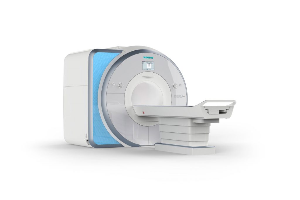
Magnetic Resonance Imaging (MRI)
Magnetic resonance imaging is a modern imaging technology that functions without the need for x-rays. Strong magnetic fields and radio waves serve to produce detailed images of the inside of the human body.
Image contrast comes about as a result of the differing responsivity of the electromagnetic waves emitted by the examined organs or tissues. High resolution image slices of the inside of the body can thereby be generated that allow determining the exact diagnosis of an illness. An MRI is therefore the ideal method to examine any part of the human body.
Main usage areas:
- Central and peripheral nervous system
- Spine and joints
- Neck, upper abdominal organs (including MRCP) and pelvic organs
- MRI mammography
- MR angiography (head and neck, thorax-abdomen-pelvis and peripheral vascular vessels)
- Study of the small and large intestines
- Urinary tract and bladder
- High-resolution multi-parametric MR imaging of the prostate
- Structural and functional diagnostics of the temporomandibular joint
- Structural and functional diagnostics of the pelvic floor (MRI defecography)
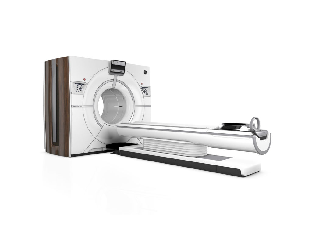
Computer Tomography (CT)
The computerised tomography is a fast and extremely precise diagnostic imaging procedure, which is able to visualise large sections of the body with detailed accuracy within seconds.
Here the body is “dissected” and visually represented by cross-sectional images of less than 1-mm thickness. Just as with standard x-rays, the different body tissues’ varying permeability to x-rays is being exploited here, too. The denser the tissue, the harder it is for x-rays to permeate through it. It is therefore possible, for example, to distinguish between bones, air (inside the lungs), water retention in the body, or to identify soft tissue. The CT is an invaluable procedure that can solve many medical queries.
Main usage areas:
- Skull, as well as thin-section CT of the petrous bone and the paranasal sinuses
- Neck (cervical spine and soft tissue)
- Thorax and HRCT of the thorax
- Abdomen and pelvis
- Skeleton
- CT angiography of all vascular territories
- CT myelography
- CT-controlled infiltration of the whole spine for pain management

Sonography/Ultrasound (US)
Sonography, also called echography or ultrasound, is the most widely used imaging method in healthcare. Due to this procedure being widely available and thanks to it lacking any radiation exposure, it may safely be used on any patient.
This makes ultrasound examinations ideal for a first assessment where the clinical picture is unclear, but also for monitoring progress. For instance, in the breast, suspected tumour findings, can be identified of a certain minimum size (approx. > 5 mm), allowing a first assessment as to their malignancy. In addition, ultrasound-controlled biopsies may be carried out.
However, physical differences do exist when it comes to the organ structures to be examined, as well as the results to be achieved in each individual patient. For example, tissue types primarily differ due to their respective condition and are either well suited for sonographic imaging or less well-suited. In general, all organs rich in blood and containing water tend to be well-suited for sonographic examination. By contrast, all gaseous organs, e.g. meteoristically distended small or large intestines, the lungs or bones, are difficult to detect by sonographic imaging. What’s more, some organs are hardly visible when healthy and normal, but become easily recognisable when diseased and enlarged (appendix, ureter, adrenal glands).

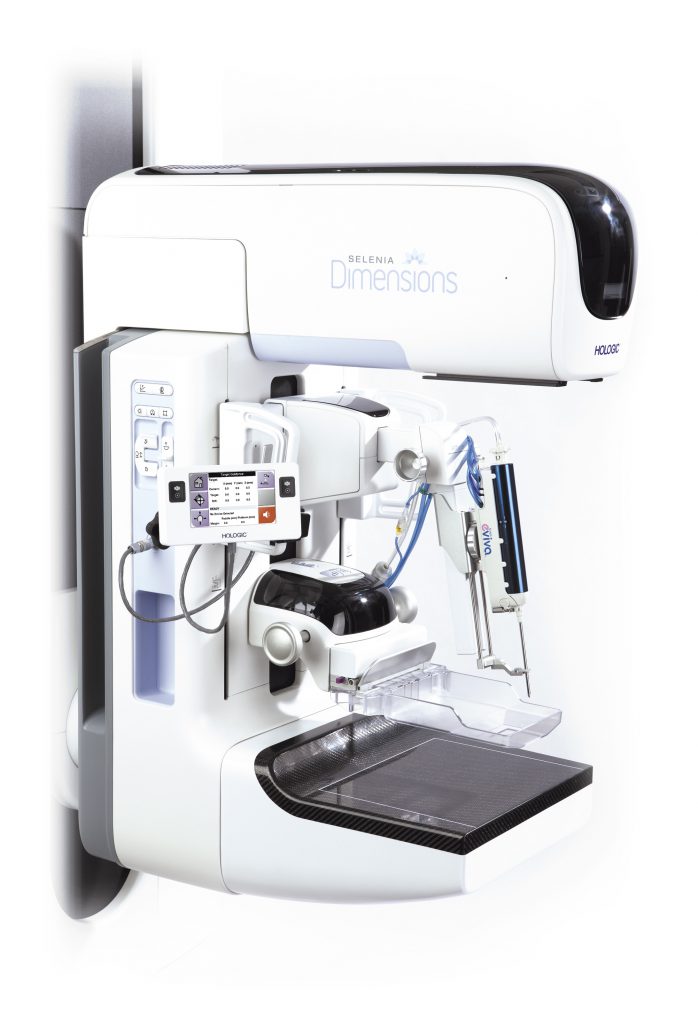
Breast Diagnostics – Mammography & Tomosynthesis
A mammography is the X-ray imaging of the breast to screen for and show benign or malignant changes. This examination is the only recognised screening method for early breast cancer detection.
In our institution we have two latest generation digital mammography systems of Hologic and Siemens.
Tomosynthesis is a modern procedure in digital mammography. It generates a series of continuous image layers throughout the whole breast, thereby enabling three-dimensional mammography imaging. It makes it possible to clearly identify structures and overlaps.
The earlier the breast cancer is detected, the better the chances of recovery and the lower the therapeutic and psychological burden on the patient.
We also perform macro-biopsies and excisions under stereotactic / echographic control. Furthermore, we actively participate in the BEJUNE breast screening programme.
Minimally Invasive Interventions and Pain Treatment
Punctures and biopsies are examinations carried out using an imaging guidance to allow sampling of suspect areas in a micro-invasive, painless and rapid manner.
Local injection therapy is a proven means of treating various complaints, especially back pain and joint problems.
A well-established method is ultrasound – and especially for the spine – spinal CT-guided infiltration therapy. It is a superior procedure thanks to the exact placement of the needle and the resulting precision of the injection site for the medication or of the biopsy. For more information please see the “Patient Information” section.
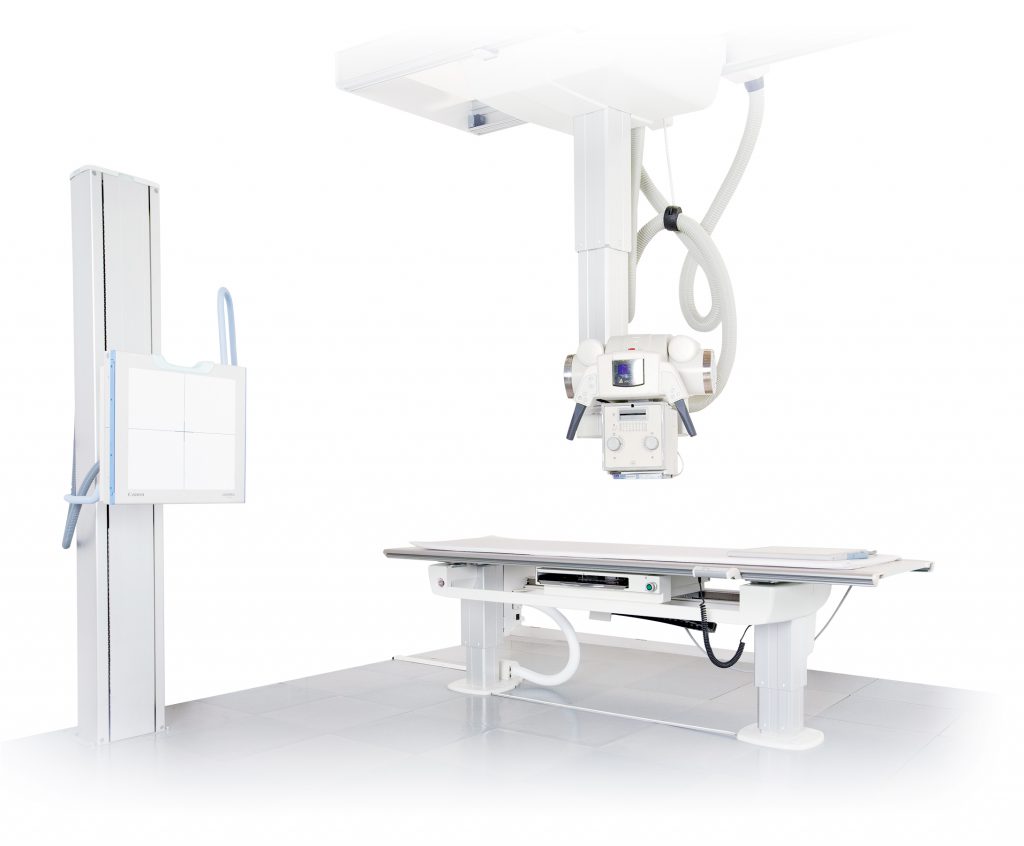
Digital Radiology – X-Ray
The classic “X-ray” to this day still forms an integral part in routine diagnostics. Frequently, x-ray images of, let’s say the lungs, of bones and joints or of the spine, are the very first step and are able to answer many questions.
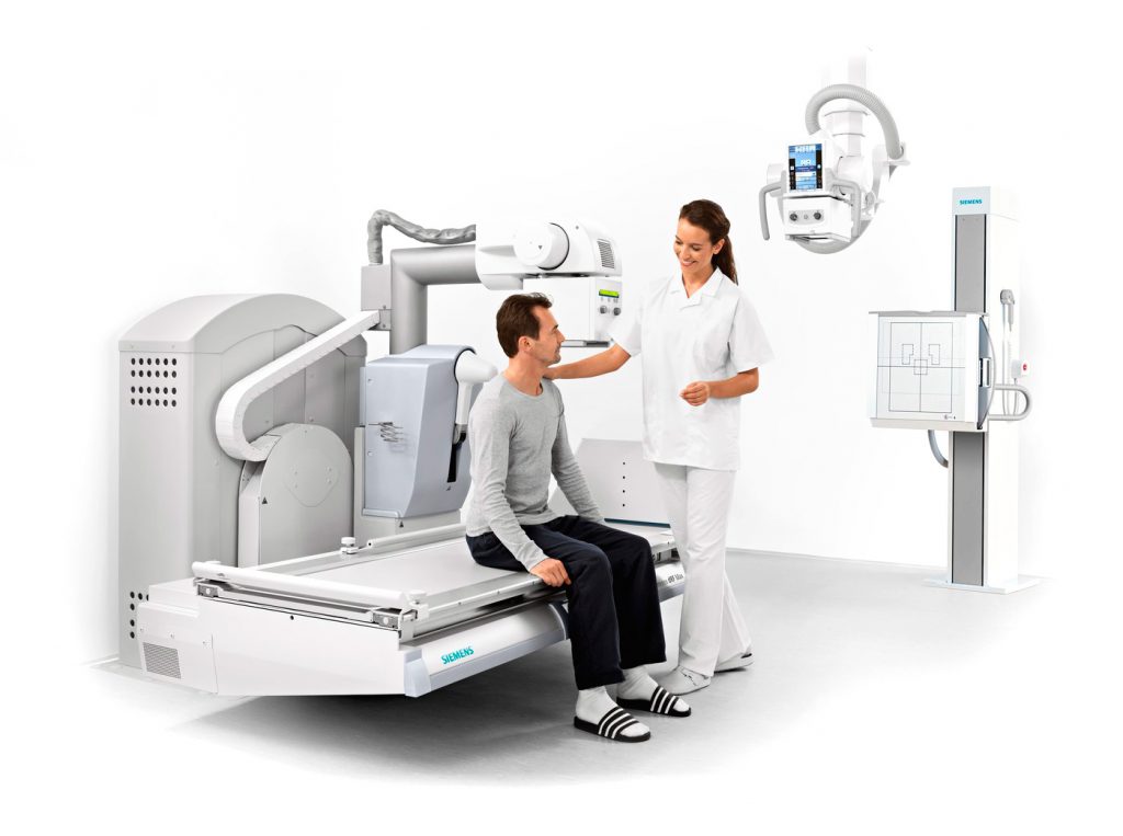
X-ray/Fluoroscopy
A fluoroscopy is often used as an additional investigation to the overview scans in order to get a better localization of pathological processes.
It is also an important part of dynamic examinations, such as in the imaging of the oesophagus, the stomach, the intestines, in arthographies (visualising the joints by injecting an iodine contrast agent), or in myelographies (visualising the spinal canal).
In this radiological screening or fluoroscopy, a continuous series of x-ray images is recorded instead of one single image (radiograph). This method also allows tracking an X-ray examination in real time on a monitor using image intensifiers. Main usage areas:
- Functional analysis of swallowing
- Oesophagus, stomach and bowel examinations
- Arthrographies of joints (shoulder, elbow, wrist, hip, knee and ankle)
- Defecography
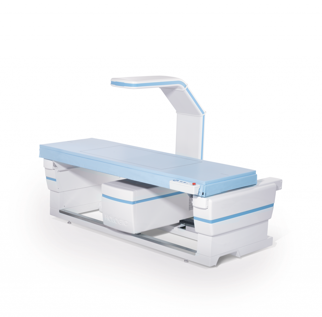
Mineralometry/Osteodensitometry (Bone Density Measurement)
Osteoporosis is a disease of the entire skeleton with reduction in bone mass and increased risk of fracture.
With the first fracture, the risk of further fractures increases. The deformed spine results in static changes that in turn often lead to chronic disorders of the locomotor system. Nowadays, the diagnosis is primarily arrived at through osteodensitometry, which is a special, low-radiation method of computerised tomography, whereby the bone density of lumbar vertebrae and that of the femoral head are measured, on which the subsequent assessment is based.